
Advance Knobology for B-Mode Imaging - Tissue Harmonics
Tissue Harmonics
Tissue Harmonic Imaging (THI) exploits non-linear propagation of US through the body tissues. Tissue is not linearly elastic, it contracts less than it expands. During tissue contraction, tissue density increases, causing the peak of the sound wave to travel slightly faster than the trough. The result of this process, called non-linear propagation, is that the wave becomes progressively more asymmetrical. This asymmetrically distorted wave is the sum of the fundamental Hz and harmonic Hz. Harmonics are higher frequency and low amplitude.
Let's illustrate this with an animation.
The transducer emits an US pulse at fundamental Hz (ideally, low Hz, high amplitude, narrow band) which hits the object at the Focal Zone. As the pulse wave travels through the tissues, the waveform distorts and generates harmonic echoes from tissue. Both fundamental and harmonic echoes return to the transducer. Ideally, the transducer is able to receive high Hz, low amplitude and broadband echoes in order to adequately filter the waves with fundamental Hz. Subsequently, only the remaining signals (mostly harmonics) are used for image generation. At present, only 2nd order harmonics are used as higher harmonics have decreasing amplitude - insufficient to generate a proper image.
The transducer emits an US pulse at fundamental Hz (ideally, low Hz, high amplitude, narrow band) which hits the object at the Focal Zone. As the pulse wave travels through the tissues, the waveform distorts and generates harmonic echoes from tissue. Both fundamental and harmonic echoes return to the transducer. Ideally, the transducer is able to receive high Hz, low amplitude and broadband echoes in order to adequately filter the waves with fundamental Hz. Subsequently, only the remaining signals (mostly harmonics) are used for image generation. At present, only 2nd order harmonics are used as higher harmonics have decreasing amplitude - insufficient to generate a proper image.
This figure maps the fundamental, harmonic, and echoes intensities in a typical US field.
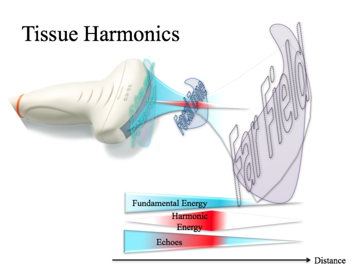
Fundamental (F): Its energy reduces away from the transducer. However, its intensity (energy per area) increases as the beam narrows (Focal Zone). As the beam advances into the Far Field, intensity reduces as energy wanes and beam profile widens.
Harmonic (H):
Its energy is primarily generated in regions where fundamental energy is intense - highest in Focal Zone. At the proximal Near Field, (eg. skin), no fundamentals are created. Beyond the Focal Zone, H generation is weak as F intensity reduces significantly. Of note, H amplitude is proportional to the F amplitude SQUARED. Hence, low amplitude F generate weak to no H.
Echoes from Harmonics:
As H are generated, they return as echoes. However, the magnitude of attenuation is proportional to the distance traveled by an echo wave. The farther it is from the transducer, the more energy lost. At the proximal Near Field, no H is generated, therefore echoes consist of only F. In the mid-Near Field, H is beginning to be produced and returned. At the Focal Zone, H are most strongly generated. Beyond the Focal Zone, H generation is weakly generated as F energy wanes, and attenuation due to the increasing distance of the reflection pathway dominates.
Nevertheless, F is more susceptible to attenuation due to distance-traveled compared to H waves. Think of it like this, to strike an object, the F wave has to travel from the transducer to the object, then the same distance back upon reflection. However, for a H wave, given it is generated and emitted from the object, the wave only has to travel from the object to the transducer - half the distance of a F wave.
Nevertheless, F is more susceptible to attenuation due to distance-traveled compared to H waves. Think of it like this, to strike an object, the F wave has to travel from the transducer to the object, then the same distance back upon reflection. However, for a H wave, given it is generated and emitted from the object, the wave only has to travel from the object to the transducer - half the distance of a F wave.
Effects:
- Improves Lateral Resolution: as harmonic beam width is narrow
- Reduces Axial Resolution: higher frequency does result in shorter spatial pulse length which should improve axial resolution. However, higher frequencies are more susceptible to attenuation. In addition, harmonic leakage (fundamental frequencies overlapping harmonic frequencies) and inadequate filtering of these frequencies will degrade axial resolution - to the extent whereby tissue planes are blurred. However, these are less problematic with the development of broadband transducers.
- Improves Contrast Resolution: increased signal-to-noise ratio results in better tissue contrast enhancement
- Improved Imaging of Deeper Tissue: generating the echo from deep structures of interest then receiving it (Harmonics) mitigates attenuation due to scatter from fat tissue
- Decreases Near Field Clutter: harmonic waves are weakly or not produced in the Near Field, therefore clutter (artifacts with weak echo amplitude which are generated in the superficial layers), THI can circumvent detection of these extraneous signal
- Reverberation Attenuation: as most reverberations are created at tissues interfaces that are relatively superficial in the human body
- Decreases Side Lobe Artifacts: low energy lobes lobes generate very weak or no harmonics, therefore the artifacts will be suppressed with THI
- Reduces the Dynamic Range
Take Home Messages:
Practical Points:
- As the beam approaches the Focal Zone, more and more harmonic energy is produced
- Paucity of harmonic echoes return from the proximal Near Field and Far Field
- Artifacts are primarily consists of fundamental frequencies, therefore when THI is On, these noise can be filtered to yield a cleared image
- Toggle between THI On and Off to see which yields the best image quality for your purposes
Practical Points:
- Move the Focus of the beam to the region of interest to maximize harmonic wave generation should you want to employ THI
- Imaging proximal Near Field with THI in a "Good US subjects" (thin individuals) usually yields poor Tissue Harmonic images compared to one generated from Fundamentals - eg.) Most often encountered when acquiring an apical view in a thin individual. The apex looks poorly defined. Turn THI Off and see if the image quality improves.
- Some artifacts are accentuated or attenuated with THI - an advantage or disadvantage depending the PoCUS Indication:
- Thoracic PoCUS for A-lines: THI attenuates reverberation, so turn THI Off
- Needle Imaging for procedure: THI attenuates the reverberation and ring down, so turn THI Off