
Pneumothorax - Sonographic Signs
Pneumothorax
Most of the sonographic features of pneumothorax are actually not foreign concepts if you have been studying the module in sequence. The only new sign is the Lung Point.
1. What are the sonographic features?
a. Pleural Sliding
b. B-Lines
c. Lung Pulse
d. Lung Point
Most of the sonographic features of pneumothorax are actually not foreign concepts if you have been studying the module in sequence. The only new sign is the Lung Point.
1. What are the sonographic features?
a. Pleural Sliding
b. B-Lines
c. Lung Pulse
d. Lung Point
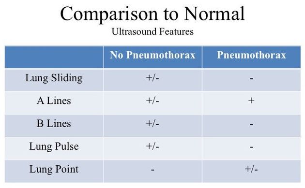
1. What are the sonographic features of pneumothorax?
- Pleural Sliding: Absent (Stratosphere sign)
- A-Lines: Present
- B-Lines: Absent
- Lung Pulse: Absent
- Lung Point: Maybe Present
a. Pleural Sliding:
Of all the pneumothorax sonographic signs, this is absolutely THE MOST IMPORTANT one to master. The reason is that this is the simplest to visualize and, if present, pneumothorax is ruled out. Unfortunately, its absence does not rule in a pneumothorax. As you may recall, pleural sliding signifies VPPI are apposed and moving against each other. However, even if you do not see sliding, VPPI can be in contact but just not moving against each other - eg. pleural adhesion.
Pleural Sliding in B-Mode:
A shimmering glitter and a to-&-fro motion are exhibited by the pleural line. These features suggest that the not only the VPPI are intact, but they are sliding against each other. This is a normal finding. When you see this, there is NO pneumothorax at the region being visualized.
Pleural Sliding in M-Mode:
It is an efficacious modality to interrogate for pleural sliding should it be difficult to discern with B-Mode. The resultant graphic divides into two portions:
- Top Portion above the pleural line: consists of tissues and muscles above the pleural line which are not moving, therefore the M-Mode records no movement over time along the sample (Green Line). Hence, that segment consists of continuous bars of back/grey/white.
- Bottom Portion below he pleural line:
below the pleural line the segment is much more grainy. This "grainy" is due to motion of the VPPI.
This picture has been described as the Sea and Sandy Beach = Pleural Sliding is PRESENT.

Pleural Sliding Absent in B-Mode:
This image was acquired from a patient with a right pneumothorax. As you may appreciate, there is NO pleural sliding present. It is completely motionless despite there is active respiration (as indicated by the movements of the intercostal muscles).
This image was acquired from a patient with a right pneumothorax. As you may appreciate, there is NO pleural sliding present. It is completely motionless despite there is active respiration (as indicated by the movements of the intercostal muscles).
A-Lines Present and Pleural Sliding Absent:
The former indicates that there is air in the thorax. This means either (a) a pneumothorax is present, or (b) there is no pneumothorax BUT the visceral pleura of an aerated lung is apposed to the parietal pleura. Hence, pneumothorax cannot be ruled out with these two signs alone.
The former indicates that there is air in the thorax. This means either (a) a pneumothorax is present, or (b) there is no pneumothorax BUT the visceral pleura of an aerated lung is apposed to the parietal pleura. Hence, pneumothorax cannot be ruled out with these two signs alone.
Is there any visible pleural sliding:
This clip was attained with the linear probe is placed parallel
to the ICS, therefore the muscle and tissue fibers are viewed longitudinally. Given this patient is recruiting accessory muscles, their motion confounds pleural movement.
Hence when assessing pleural sliding, it is preferable to have the probe perpendicular
to the ICS.
M-Mode of a pneumothorax (left half) and a normal lung (right half) are placed side by side for comparison.

The "Sea" component of a pneumothroax is the same as a normal thorax given that there is no movement in this region. However, the "Sandy Beach" component is not present in a pneumothorax. That grainy appearance is substituted by a barcode-like pattern given that there is NO movement at the VPPI.
This pan-barcode appearance has been termed the Stratosphere Sign which indicates there is NO pleural sliding. However, the VPPI could potentially be apposed, therefore this sign cannot rule in pneumothorax.
This pan-barcode appearance has been termed the Stratosphere Sign which indicates there is NO pleural sliding. However, the VPPI could potentially be apposed, therefore this sign cannot rule in pneumothorax.
b. Lung Pulse: refers to the Pleural Line moving in a pusatile, rhythmic manner with a rate that is consistent with the cardiac pulsation. The reason why this is visible at the Pleural Line is because when the VPPI are apposed, the motion transmitted from the heart to the visceral pleura can be imaged with US. However,when the VPPI is separated by either air (or fluid), the visceral pleural pulsation cannot be imaged. This is sign is best seen in Z1-2 and tend to be attenuated at Z3-6 due to distance from the heart. Its presence rules out a pneumothorax, but absence does cannot rule in.
Lung Pulse Present in B-Mode:
This video clip demonstrate the Lung Pulse in a normal thorax. For ease of visualization, the subject was asked to breath-hold so that active respiratory movements will not obscure this subtle finding. Note the rate and rhythmic movement of the Pleural Line reflects the underlying heart rate and normal sinus rhythm of the subject.
Lung Pulse Present in M-Mode:
Manifests as the rhythmic vertical perturbation that TERMINATE at the Pleural Line. This is vital. Any column extending superior to the Pleural Line is NOT Lung Pulse.

Lung Pulse Absent in B-Mode:
In this clip, one can clearly identify a Pleural Line that is stationary with A-lines deep to it. This means the VPPI is either apposed and not moving against each other, or the VPPI is separated. If the former is true, one expects to see Lung Pulse. However, in this case, there is NO rhythmic pulsation - no visible lung pulse. Unfortunately, due to this sign's limitations, its absence cannot rule in a pneumothorax (not sufficiently specific).
Lung Pulse Absent in M-Mode:
Stratosphere Sign is seen with visible vertical columns. However, notice how they all extend superior to the Pleural Line. These are NOT Lung Pulses, but are motion artifacts created SUPERIOR to the VPPI - eg.) Probe, muscle movements.

c. B-Lines:
refers to the artifacts that arises from the Pleural Line and extend vertically down to the edge of the screen and moves with respiration. The key point is that B-lines can only be generated when VPPI are apposed.
Consequently, if this artifact is present, there is NO pneumothorax in the region being imaged. Unfortunately, like the signs above, its absence CANNOT rule in pneumothorax.
Z-lines have been found to present in both normal thoraces and pneumothoraces, therefore it cannot be used to rule out a pneumothorax.
Z-lines have been found to present in both normal thoraces and pneumothoraces, therefore it cannot be used to rule out a pneumothorax.
d. Lung Point:
refers to when the interface between the pneumothorax and where the lung parenchyma that still contacts the parietal pleura moves into view of the ultrasound beam with respiration. Given this premise, the lung point is the most specific sign for pneumothorax. However, one can imagine this interface, the Lung Point, is not easy to locate. Also, if the pneumothorax is too large, no visceral pleura will come in contact the parietal pleura, therefore the Lung Point cannot be imaged. As a result, the this sign's sensitivity is quite poor.
Lung Point:
The probe is imaging where the border between the pneumothorax and a region where the VPPI are in contact (visceral pleura is contact the parietal pleura) - as illustrated by the schematic diagram.
Whenever the patient inspires, the apposed VPPI move into the pneumothorax and into view of the tranducer's FOV. Hence, one sees a structure slides into view (then out with expiration).
This structure is most likely the lung though it could be the heart that you are seeing - will be discussing this in the Pitfalls section.

Lung Point on M-Mode: It manifests as intermittent interruption of the Stratosphere by the Sandy-Beach.
In order to attain this pattern, however, you have to ensure that the M-Mode sampling gate (Green Line) crosses the segment of the Pleural Line that the Lung Point will definitely slide into.
In order to attain this pattern, however, you have to ensure that the M-Mode sampling gate (Green Line) crosses the segment of the Pleural Line that the Lung Point will definitely slide into.

The Lung Point is not easy to find. You hope to be able to find that apposed VPPI as you slide the probe laterally from the peristernal line.
However, you can image that a pneumothorax can become so sizeable such that the entire atelectatic lung is completely surround by air with no visceral pleura contacting the parietal pleura - as illustrated. Hence, the Lung Point cannot be elicited under such circumstances.
However, you can image that a pneumothorax can become so sizeable such that the entire atelectatic lung is completely surround by air with no visceral pleura contacting the parietal pleura - as illustrated. Hence, the Lung Point cannot be elicited under such circumstances.
Take Home Messages:
- Presence of Pleural Sliding, B-Lines, or Lung Pulse indicates VPPI are apposed = NO pneumothorax in the region being scanned
- Absence of Pleural Sliding, B-Lines, or Lung Pulse are not sufficiently specific to rule in pneumothorax - other thoracic pathologies can, also, exhibit the absence of these signs
- Presence of Lung Point = Pneumothroax
- Absence of Lung Point does NOT rule out pneumothorax