
Hydronephrosis - Sonographic Features
1. Normal
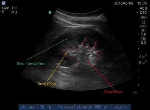
Right kidney in Long Axis:
In a normal kidney, the renal pelvis is uniformly echoic relative to the parenchyma.
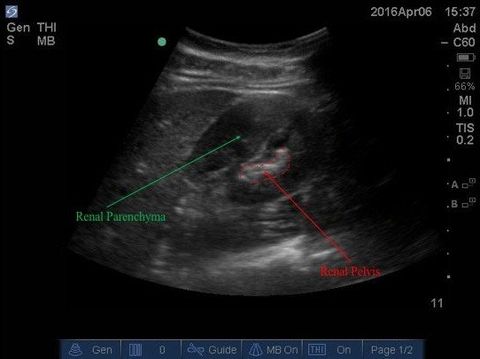
Right kidney in Short Axis:
In a normal kidney, the renal pelvis is uniformly echoic relative to the parenchyma.
2. Hydronephrosis
Hydronephrosis:
(1) the renal pelvis and the calices are hypoechoic/anechoic - signifying dilatation; & (2) these regions are interconnected to the renal pelvis -- both criteria are vital.
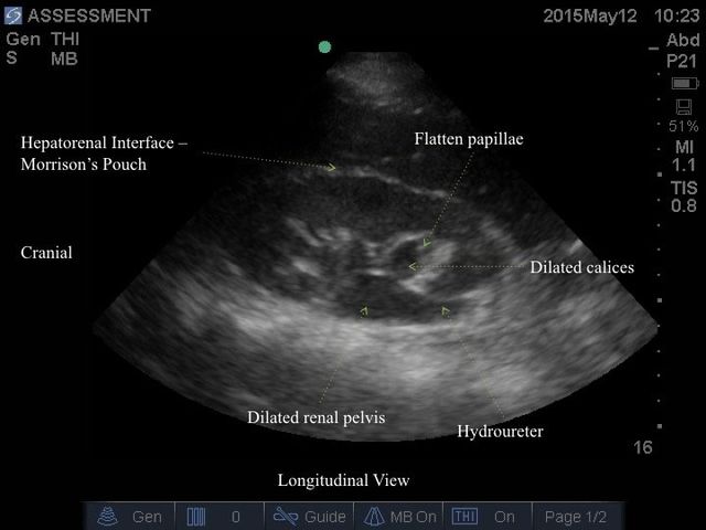
Short axis view of the same case. Take note of the dilated calices drain into the dilated renal pelvis.
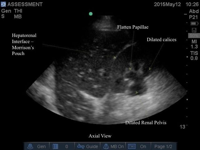
A case of severe hydronephrosis with a massively dilated pelvis, calices, and flattened parenchyma. Note, there is a renal cyst a about 11am (it does not connect to the renal pelvis).
Take Home Message:
- If there is anechoic or hypoechoic regions within the renal pelvis, trace them to see if they drain into the renal pelvis
- Be wary of renal cyst as a confounder -- it does NOT connect to the renal pelvis