
Pericardial Effusion - Pitfalls
1. Pericardial Fat Pad
This is the MOST
common mimic of pericardial effusion. It is a piece of adipose tissue that could be on the epicardium or on pericardium itself.
It is usually seen in the anterior (parasternal long/short views) and inferior aspects of the heart (subcostal view).
Its appearance is speckled AND moves synchronously with the cardiac motions.
Here we have a parasternal long view. Notice just anterior (above) to the LV and the RVOT, there is a speckled layer.
Also, notice how the speckles' direction of motion is synchronous to the myocardial systolic and diastolic movements.
This is the pericardial fat pad.
There is, also, a fat pad here though much thinner than previous and the speckles are not as obvious.
Hence, it maybe mistaken as a pericardial effusion.
However, if you magnify the region, you will notice the speckle. Nevertheless, if it is hard to discern, it is vital to remember to confirm what you saw with at least two views.
The pericardial fat pad can be quite difficult to visualize due to its similar echogenicity with the adjacent cardiac structures.
Here, the presence of an anechoic pericardial effusion contrasts the fat pad. Notice it is this "flabby" piece of echoic and speckled tissue that is adhered to the epicardium.
2. Other "Effusions"
Pleural effusions or
ascites can mimic a pericardial effusion.
Left Pleural Effusion Mimic
Given the heart is positioned in the left thorax, fluid collection around the heart and be confounded by a left pleura effusion.
Notice there is an anechoic collection just posterior (far field) to the heart.
It looks like a pericardial effusion. However, this is, in fact, a pleural effusion. It is the left pleural effusion to be exact.
To distinguish between pericardial effusion vs pleural effusion in the parasternal long view, one can use the descending aorta as the landmark.
The pericardial effusion tracks anterior to the descending aorta; and the pleural effusion tracks posterior to the descending aorta.
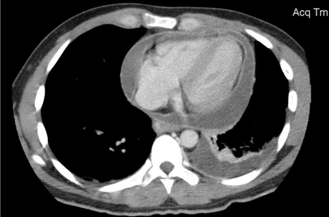
To understand how the fluid tracts in front and behind the descending aorta can be of help distinguish a pericardial effusion from a left pleural effusion, take a look at this cross-section CT here with both.
Notice where the aorta is in relation to the two fluid collection.
Here we have a clip where a left pleural effusion and pericardial effusion are both present taken from the parasternal long view.
The pleural effusion tracks posterior to the descending aorta.
The pericardial effusion tracks anterior to the descending aorta.
Right Pleural Effusion Mimic
This is not as commonly seen, but it can be quite the confounder.
Please take a look at the clips posted by Dr. Virginia Zarama (Twitter: @VirginiaZarama) on https://twitter.com/VirginiaZarama/status/1205700600979247104?s=20
Ascites Mimic
This
is a common problem when imaging from a subcostal view. The best way to mitigate this problem is, well, image the heart from other views.
Here we have a subcostal view. You can see the RV and the LV here. Notice there is an anechoic fluid collection there. One may think this is a pericardial effusion.
However, it is EXTERNAL to the pericardium (hyperechoic sac). This is, in fact, ascites.
You can see the liver coming in-and-out of view. That small strand is the faciform ligament.
Here is another ascites mimic taken from the subcostal view.
The pericardial scan is not as obvious this time, but can still be distinguished. Notice the tricuspid annulus is moving independent to the surround white line which is the pericardium.
The best way is to clarify this issue is to image the heart from other views.
Take Home Messages:
- Pericardial fat pad has speckles that move synchronously with the heart
- Pericardial effusion tracks anterior to the ascending aorta
- Image the heart from more than one view to verify the presence of a pericardial effusion