
Ascites and Paracentesis - Sonographic Findings
Ascites
1. Hepatorenal Space
2. Splenorenal Space
3. Ascites Pocket
4. Bladder
5. Superficial Vessels
6. Fluid Appearance and Content
Sonographic Features of Ascites PoCUS:
2. Splenorenal Space
3. Ascites Pocket
4. Bladder
5. Superficial Vessels
6. Fluid Appearance and Content
1.
Hepatorenal Space (aka. Morrison's Pouch)
This potential space between the liver and right kidney is relatively easy to see given the size of the liver. When fluid collects here, the space will be separated and occupied by either anechoic/echoic fluid.
This potential space between the liver and right kidney is relatively easy to see given the size of the liver. When fluid collects here, the space will be separated and occupied by either anechoic/echoic fluid.
2. Splenorenal Space
This potential space between the spleen and left kidney is more difficult to image as the spleen is smaller and more posterior than the liver, therefore the acoustic window is much smaller. When fluid collects here, the space will be separated and occupied by either anechoic/echoic fluid.
This potential space between the spleen and left kidney is more difficult to image as the spleen is smaller and more posterior than the liver, therefore the acoustic window is much smaller. When fluid collects here, the space will be separated and occupied by either anechoic/echoic fluid.
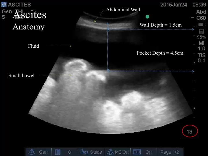
3. Ascites Pocket
This pocket of peritoneal fluid collection was imaged at the right lower quadrant. The abdominal wall and fluid pocket can be seen. The critical structure in this view is the small intestine - circular/ovoid and echogenic. Sometimes, the contents of the bowel can be seen if there is no air within.
This pocket of peritoneal fluid collection was imaged at the right lower quadrant. The abdominal wall and fluid pocket can be seen. The critical structure in this view is the small intestine - circular/ovoid and echogenic. Sometimes, the contents of the bowel can be seen if there is no air within.
4. Bladder
There are other critical fluid filled critical structures in the abdomen. The bladder is the most common structure encountered especially at the suprapubic region. A large ovarian cyst is, also, not uncommon
The key message here is that whenever you see fluid, look around for walls containing that fluid. If the fluid is contained, see if you can identify the structure. However, if you are ever in doubt, obtain definitive imaging.
5. Superficial Vessels
Using the linear probe, one can assess the potential abdominal wall region for paracentesis for underlying blood vessel. If a vessel is, indeed, present, avoid that spot lest laceration of the vessel resulting in hemorrhage.
Using the linear probe, one can assess the potential abdominal wall region for paracentesis for underlying blood vessel. If a vessel is, indeed, present, avoid that spot lest laceration of the vessel resulting in hemorrhage.
6. Fluid Content
Figure i. Anechoic and Clear Ascites:
This image demonstrate Black looking fluid in the peritoneal cavity with no evidence of "debris" in it. Some of the hazziness adjacent to the bowel are due to artifact.

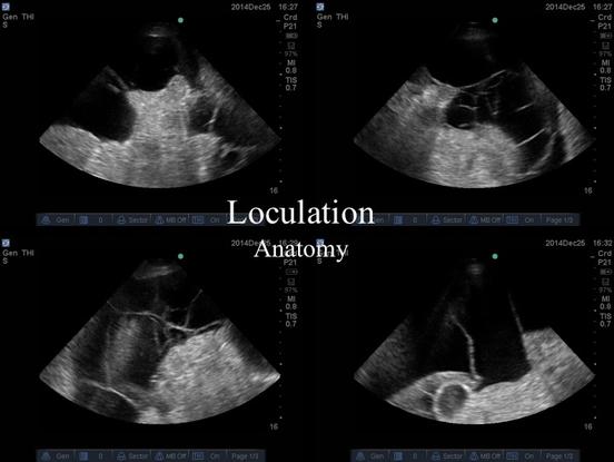
Figure ii. Loculations: These four images show septae and fibrin strands attached to the bowel contents. Some are even anchoring at the peritoneum at their terminal ends. These are intra-peritoneal space can be very complex with intricate loculations.
Clip iii. Turbid Ascites:
On initial glance, you can tell there is an anechoic. However, looking at the dependent region of the pocket, there are echogenic particulates that are swirling accordingly to the forces applied to the fluid - be it the diaphragmatic motion or bouncing the probe up and down on the abdominal wall.
As seen on Figure i, some hazzy artifact is sometime seen adjacent to the bowel. This hazziness can look like particulate matter as well. However, the artifacts DO NOT swirl accordingly to changes in intra-abdominal forces.
As seen on Figure i, some hazzy artifact is sometime seen adjacent to the bowel. This hazziness can look like particulate matter as well. However, the artifacts DO NOT swirl accordingly to changes in intra-abdominal forces.
Take Home Messages:
- Screen for intra-abdominal fluid at the hepatorenal and splenorenal spaces
- When imaging for a site for paracentesis
- scan the regions that are clearly "bulging" - usually at RLQ, LLQ, and Flank
- avoid suparpubic and least dependent regions
- look for an "Ideal" paracentesis site - safe spot