
The processor displays the transduced electrical signals from the probe with a spectrum of brightness on the monitor.
The brightness displayed corresponds to the amplitude of the echo. In addition, B-Mode yields 2 dimensional images in real-time, therefore a motion can be captured.
All machines default to this mode when the machine is turned on.
On the Sonosite-M Turbo, it is the "2D" button, and on the TE7, it is the "B" button.

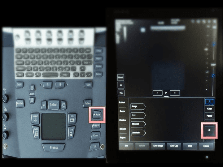
Another basic imaging mode to familiarize with is the M-mode,
or Motion—mode. When this modality is selected, only one sliver or one dimension on the B-mode image stemming from the probe footprint will be displayed on the monitor over a time axis.
On the screen, it produces a graph of what is happening to all the structures along a "line of sight" over time. In this case, this "line of sight" is indicated by the red arrow. Here we have the parasternal long access of the heart with the "line of sight" moved to cross the mitral valve.
It is very helpful for motion analysis and measuring distance/depth of structures.
On the Sonosite-M Turbo, it is the "M Mode" button, and on the TE7, it is the "M" button.
2. Gain: it is the amplification of the transducer's electrical signal to produce an image
So let's take a look at it graphically. The heart is to be ultrasounded. The transducer sends a pulse wave striking the object of interest (ie. heart). An echo is generated from that object and reflected back to the transducer. The transducer converts that sound signal to an electrical signal which is then relayed to the processor. Subsequently, the processor amplifies the electrical signal and display it as an image on the monitor.

When you are changing the Gain via the Interface, you are basically manipulating how much amplification the processor will yield.
If you increase the Gain, you will notice the image wll get brighter overall.
If you reduce the Gain, the image will become dimmer and dimmer.

What is the Optimal Gain?
Too much: the image is too bright; and Too little: the image is too dark. Either would result in information being masked by the unregulated amplification process.
Practical Point:

Here is a video of demonstrating Gain manipulation.
Take note of what happens to the definition of the sternocleidomuscle, vessels, and the surrounding tissues as the Gain is changed.
Like the Gain function, you can have too much depth. The Optimal Depth is when the structure(s) of interest is shown in its entirety and displayed in a reasonable portion of the monitor. This concept is called the Visual Real-Estate. You want to maximize the VRE of structure(s) of interest.
In this case, you want to image the carotid. On the upper left, the carotid is seen, but occupies very little of the VRE, therefore more finite features cannot be appreciated. Too little depth, you only see part of the carotid. With the optimal, you can see the entire carotid in detail.

The Depth control is enveloped in the Red Squre on the Sonosite M-Turbo and Mindray TE7.

- B-Mode Imaging: 2D imaging over time
- M-Mode Imaging:1D imaging over time
- Gain:amplification of echo signals - optimal when the structure of interest is seen and well defined
- Depth:vertical distance to be displayed on the screen - optimal when visual real-estate is maximized