
Case 4 - Post-Thoracentesis
Indication:
Given the examination findings, it was most likely bilateral pleural effusion. The use of the PoCUS was to verify the presence of the effusion, size, and feasibility for thoracentesis.
Image Acquisition:
Given the examination findings, it was most likely bilateral pleural effusion. The use of the PoCUS was to verify the presence of the effusion, size, and feasibility for thoracentesis.
Image Acquisition:
The patient was laid flat on the bed with the probe placed at the most independent point on the thorax, between the peristernal and mid-clavicular line, and perpendicular to the rib space. The probe was then translated from the highest point in the thorax to its lowest point along this defined zone to interrogate for lung sliding.
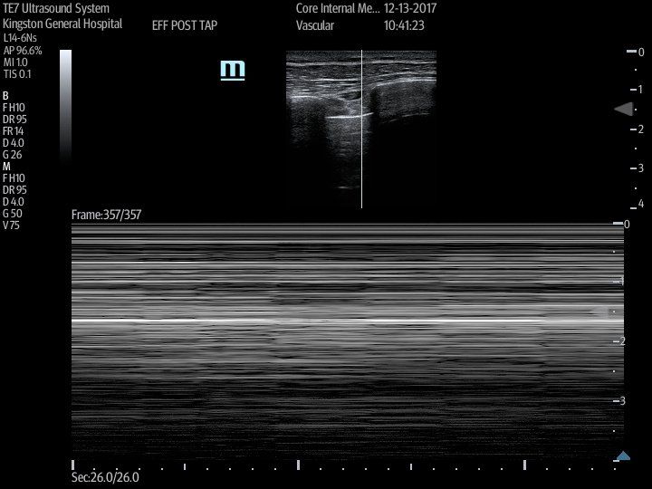
Image Acquisition:
Given the above findings, the concern is for an iatrogenic pneumothorax. In order to verify its presence, the lung point was sought by sliding the probe laterally along the 5th ICS.
Clinical Synthesis:
The presence of the pneumothorax was detected, therefore a CXR was ordered to elucidate its size to determine whether a chest-tube is warranted.