
Advance Knobology for B-Mode Imaging - Compound Imaging
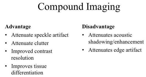
Compound Imaging
Compound Imaging:
In this modality, the US beam is steered from angle theta-max to theta-min (an arc). Subsequently, the processor averages the overlapped (compounded) regions together. (It is kind of like what HD cameras do when it takes multiple pictures and combines them into one “high” quality picture).
The processing depends on proprietary software. If an echo is consistently captured throughout all frames, the echo signal will be retained and displayed on the monitor. However, if it is not, the echo will be discarded.
The processing depends on proprietary software. If an echo is consistently captured throughout all frames, the echo signal will be retained and displayed on the monitor. However, if it is not, the echo will be discarded.
The compounded region is the purple triangular zone. The size of this region is dependent on the width of the transducer marked by the red line and angle of the steer (range of the arc).
Below are two sets of images of the cervical vessels (upper row) and right upper quadrant (lower row) taken with the Compound Imaging On and Off. Notice any differences?

Well, for the cervical vessel picture, the tissues are better defined. Not only that, take a look at the edge artifact and acoustic enhancement artifacts originating from the carotid artery, with compound imaging, it is attenuated.
How about the right upper quadrant images? Not too much changes.
How about the right upper quadrant images? Not too much changes.
Should we use Compound Imaging all the time?
The answer is NO. It has its disadvantages.
Compound imaging requires increased recording time in order to attain the information from multiple angles. Frame rate will be reduced compared to conventional imaging especially with structures that are moving rapidly.
The answer is NO. It has its disadvantages.
Compound imaging requires increased recording time in order to attain the information from multiple angles. Frame rate will be reduced compared to conventional imaging especially with structures that are moving rapidly.
Take Home Messages:
- It may improve overall image quality
- Some artifacts maybe attenuated which could be an advantage, or disadvantage (if artifact interpretation is an integral to the diagnosis)
- Turn On/Off and use the mode that yields the optimal image