
Thoracic Basics - Normal Findings
Normal Findings
1. Pleural Sliding and A-Lines
2. Z-Lines
3. Lung Pulse
4. Base of the Thorax:
Before we discuss sonographic features of pathologies, it is crucial one becomes familiar with the Normal Thoracic Findings:
2. Z-Lines
3. Lung Pulse
4. Base of the Thorax:
Normal Findings:
Pleural Sliding, A-Lines, Z-Lines, Lung Pulse can be seen almost any thoracic scanning zones. However, the Curtain, Mirror, and Spine Signs specifically refer to the sonographic findings in Zone 4.
Pleural Sliding, A-Lines, Z-Lines, Lung Pulse can be seen almost any thoracic scanning zones. However, the Curtain, Mirror, and Spine Signs specifically refer to the sonographic findings in Zone 4.
1. Pleural Sliding and A-Lines
Pleural Sliding: when the visceral & parietal pleura are apposed AND moving against each other, they manifest as a shimmering/moving hyperechoic line (pleural line). The visceral and parietal pleura cannot be resolved into separate lines via US.
A-Lines: reverberation artifacts originating from the pleural line. If you look carefully, these lines look exactly like the pleural line except they are increasingly attenuated with increasing depth. In addition, they are equi-distanced from each other.
A-Lines indicates there is air in the thoracic region being scanned - either pneumothorax vs no pneumothorax + air in parenchyma.
2. Z-Lines
This is, also, an sonographic artifact that is characterized by vertical lines that originate at the pleural line. They do not reach the inferior portion of the screen - tend to be quite short, and they do not move with respiration.
This is, also, an sonographic artifact that is characterized by vertical lines that originate at the pleural line. They do not reach the inferior portion of the screen - tend to be quite short, and they do not move with respiration.
This artifact has no clinical meaning - it can occur when the VPPI is apposed or separated (eg. pneumothorax).
3. Lung Pulse
Given the lungs are in contact with the heart, the rhythmic cardiac pulsation can be detected with US if the VPPI is apposed. The pulsation from the heart first transmits to the lung. Then, via VPPI contact, it is propagated to the parietal pleura.
Given the lungs are in contact with the heart, the rhythmic cardiac pulsation can be detected with US if the VPPI is apposed. The pulsation from the heart first transmits to the lung. Then, via VPPI contact, it is propagated to the parietal pleura.
It is difficult to see with active respiration. However, during breath hold, the pulsatility of the pleural line is easily visible particularly at Z1-2. If you time the Lung Pulse with an arterial pulse, you will notice they are synchronous.
Sometimes it is not easy to see the Lung Pulse with B-Mode. M-Mode can scrutinize for this finding.
The clip shows what you will see if Lung Pulse is present on M-Mode: two segments. The top consist of the muscles and tissues that are not moving (which yields a bar-code like pattern). The bottom portion is grainy (so-called "Sandy Beach") which suggests motion. The border between the two is the pleural line.
The clip shows what you will see if Lung Pulse is present on M-Mode: two segments. The top consist of the muscles and tissues that are not moving (which yields a bar-code like pattern). The bottom portion is grainy (so-called "Sandy Beach") which suggests motion. The border between the two is the pleural line.
Notice there are vertical columns spanning from the pleural line to the bottom of the screen? These are the lung pulses - the pulsation of the heart is transmitted all the way to the parietal pleura via an apposed VPPI. These vertical columns are synchronous with the heart, and they DO NOT cross into the bar-code zone.
The lung pulse is a bit of an abstract concept. Hopefully, this video will make it easier for you to understand this.
The video on the right shows an atelectatic lung pulsating synchronously to the cardiac (rhythmic) pulsation in a pleural effusion.
The video on the right shows an atelectatic lung pulsating synchronously to the cardiac (rhythmic) pulsation in a pleural effusion.
4. Base of the Thorax (Zone 4)
At zone 4, the presence of the diaphragm, a highly reflective specular surface, and other adjacent structures give rise to three sonographic findings relevant for assessing the base of the thorax.
a. Curtain Sign
b. Mirror Artifact
At zone 4, the presence of the diaphragm, a highly reflective specular surface, and other adjacent structures give rise to three sonographic findings relevant for assessing the base of the thorax.
a. Curtain Sign
b. Mirror Artifact
c. Spine Sign
a. Curtain Sign
This is simply the aerated lung moving into the FOV, just like a curtain descending, obscuring intra-abdominal structures intermittently in accordance to inspiration (curtain descending) and expiration (curtain ascending).
This is simply the aerated lung moving into the FOV, just like a curtain descending, obscuring intra-abdominal structures intermittently in accordance to inspiration (curtain descending) and expiration (curtain ascending).
b. Mirror Artifact
When there is only air in the thoracic region at Z4, this artifact is generated. Notice in this clip the spleen's ovid echoic structure is seen on the opposite side of the diaphragm - a Mirror Image. In reality, it does NOT exist in the thoracic cavity. This artifact, however, would be obliterated if this region of thorax consists of fluid or solid.
This animation shows you how this artifact is generated.
In practice, this particular sign is not that easy to visualize.
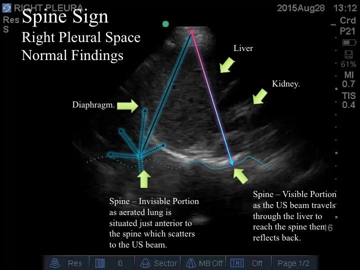
c. Spine Sign
Unlike the Mirror Artifact, the Spine Sign is not an artifact, but it, also, serves to detect the presence of fluid or solid in Z4 - in fact, it has better operational characteristics.
If there is air in Z4, the US pulse cannot reach the spine cranial to the diaphragm. As a result, that portion of the spine CANNOT be imaged. The portion that is caudal to the diaphragm can be visualized as the solid intra-abdominal organs serve as a sonographic window.
If there is fluid or solid Z4, these medium will be the sonographic conduit to the spine cranial to the diaphragm.
Take Home Message:
- Know the normal thoracic sonographic finding
- Pleural Sliding = VPPI is apposed AND moving against each other
- A-Lines = Air in the thorax
- A-Line + NO Pleural Sliding = POTENTIALLY (not definitive) a pneumothorax
- A-Line + Pleural Sliding = No pneumothorax + normal aerated parenchyma = normal region
- Z-Line
= no clinical meaning
- Mirror Artifact:
- Visible = Air in Z4
- Not visible = Fluid or solid in Z4
- Spine Sign:
- Spine not visible caudal to the diaphragm = Air in Z4
- Spine visible cranial to the diaphragm = Fluid or solid in Z4