
IVC - Image Interpretation
1. Structures
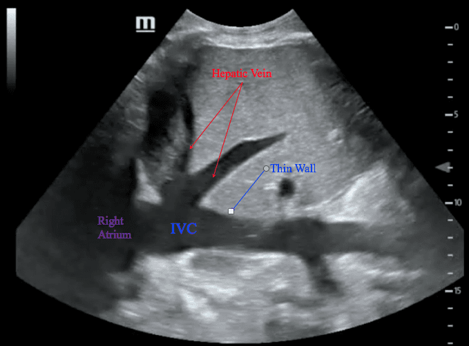
The only landmarks to verify you are indeed imaging the IVC is that (a) the tubular structure is connected to the right atrium, or (b) the hepatic vein(s).
2. Aorta
Ensure you are NOT looking at the aorta -- they are close by each other. Often it is falsely visualized as the probe is a bit towards the left of the patient.
The following video clip initially images the aorta where the probe was just left to the midline. As the sonographer slides the probe towards the right, the IVC is revealed.
Features of IVC:
- thin walled
- surrounded by liver parenchyma on both sides
- drains into RA
- hepatic vein drains into it
Features of Aorta:
- thick wall
- liver parenchyma is not around it
- not connected to the RA
- pusatile - monophasic
3. IVC Diameter
This measurement is taken at the end expiration.
It is imperative that the ultrasound beam is cutting the true diameter of the IVC longitudinally. Otherwise, it will be underestimated. See the animation below:
This measurement is taken at the end expiration.
It is imperative that the ultrasound beam is cutting the true diameter of the IVC longitudinally. Otherwise, it will be underestimated. See the animation below:
The normal diameter of the IVC is difficult to define.
American Society of Echocardiography and European Associated of Echocardiography 2011 Guidelines standardized the IVC diameter interpretation:
American Society of Echocardiography and European Associated of Echocardiography 2011 Guidelines standardized the IVC diameter interpretation:
- <1.2cm: small
- 1.2-1.7cm: normal
- 1.8-2.5cm: dilated
- >2.5: markedly dilated
Clinically, IVC size is not as useful as IVC collapsibility. With a wide "grey zone", its utility is diminished. However, at its extremes, <1cm or >2.5cm, it is more specific.
Dilated IVC:
>2.5cm

Narrow IVC:
<1cm
4. IVC Collapsibility
For this parameter, the goal is to evaluate the IVC's diameter variation during the respiratory cycle. Generally:
For this parameter, the goal is to evaluate the IVC's diameter variation during the respiratory cycle. Generally:
- Spontaneous Breathing: IVC tends to collapse during inspiration
- Mechanical Positive Pressure Ventilation: IVC tends to distend during inspiratory phase
- Spontaneous Breathing + NIPPV: difficult to predict
Collapsibility is defined by:
Collapsibility = (IVC Max diameter – IVC Min diameter)
x 100%
(IVC Max Diameter)
*Again, beware of measuring the FALSE DIAMETER -- results in false interpretation.
Reference Point along the IVC for Measuring Collapsibility:
by convention, it is set at 2cm distal to the right atrium-IVC junction as this has had been the standardized location in IVC studies.
Example:
The patient's IVC was imaged in 2D mode. Subsequently, the patient instructed to perform a rapid sniff. One can see that the IVC was collapsing during each of the inspiratory maneuvers. The degree of collapse can be "eye-balled" -- <50%.
The patient's IVC was imaged in 2D mode. Subsequently, the patient instructed to perform a rapid sniff. One can see that the IVC was collapsing during each of the inspiratory maneuvers. The degree of collapse can be "eye-balled" -- <50%.
You may, also, place a M-mode line across the IVC for a more accurate measurement of collapsibility.
Take Home Messages:
- The IVC joins into the right atrium
- The hepatic veins drain into the IVC
- Beware of measuring the false diameter of the IVC in its longitudinal view