
Pneumothorax - Pitfalls
Pitfalls
1. E-Lines ≠ A-Line
2. Lung Pulse Mimic ≠ Lung Pulse
3. Cardiac Point ≠ Lung Point
4. Hydropneumothorax
There are several noteworthy caveats to pneumothorax imaging that must be discussed to reduce interpretation errors.
2. Lung Pulse Mimic ≠ Lung Pulse
3. Cardiac Point ≠ Lung Point
4. Hydropneumothorax
1. E-Lines ≠ A-Lines
E-lines are reverberation artifacts that look like A-lines. However, E-lines arise ABOVE the Pleural Line. Most commonly, subcutaneous emphysema is the cause of the E-line artifacts.
There are several solutions to circumvent a misdiagnosis:
E-lines are reverberation artifacts that look like A-lines. However, E-lines arise ABOVE the Pleural Line. Most commonly, subcutaneous emphysema is the cause of the E-line artifacts.
There are several solutions to circumvent a misdiagnosis:
- Pleural Line is ALWAYS DEEP to the ribs of the Bat-Wing Sign
- Pleural Line is NOT that superficial/shallow in depth
- Subcutaneous air maybe displaced with pressure
2. Lung Pulse Mimic ≠ Lung Pulse
Here is another pitfall which can be quite subtle. The lung pulse is the transmission of the cardiac oscillation to the lung which is seen when there is direct contact between the visceral and the parietal pleura.
Here is another pitfall which can be quite subtle. The lung pulse is the transmission of the cardiac oscillation to the lung which is seen when there is direct contact between the visceral and the parietal pleura.

In this image, clearly there is lung pulse demarcated by the green lines here, but notice how it terminates at the pleural line. Remember that the lung pulse terminates at the pleural line. However, take a look at this image, there are these vertical columns that projects above the pleural line. These vertical lines are NOT lung pulse. They are generated from ABOVE the pleura. In this case, you can see the vertical lines spans all the way up to the top of the M mode spread – right to the where the probe contacts the skin. In fact, this is a probe pulse. This is generated when pressure is applied towards the thorax wall with the probe.
Here is a M-Mode spread taken from a patient with a hydropneumothorax. You can see the Stratosphere and the Sandy Beach alternating intermittently.
Two vertical columns are demarcated for you in Red. One spans from the skin to the bottom of the spread. The other once starts in the middle of the upper segment. Both of these are NOT Lung Pulse.

3. Cardiac Point ≠ Lung Point
When the probe is placed close to the heart to evaluate its adjacent VPPI, the heart can be seen moving in the field of view. This is usually seen in LZ2.
As you can see from this video clip, it would seem like there is a lung point present. However, notice the rate and rhythm of the movement. It is too fast and regular for respiration.
If you time it with the pulse, you will find it synchronous with the movement you are seeing on the screen. Also, there seems to be something contracting underneath. If we increase the depth, you can see the heart contracting there.
As an aside, you can tell from this clip, however, the heart is not moving into a pneumothorax region as there is a small Z-line present.
As an aside, you can tell from this clip, however, the heart is not moving into a pneumothorax region as there is a small Z-line present.
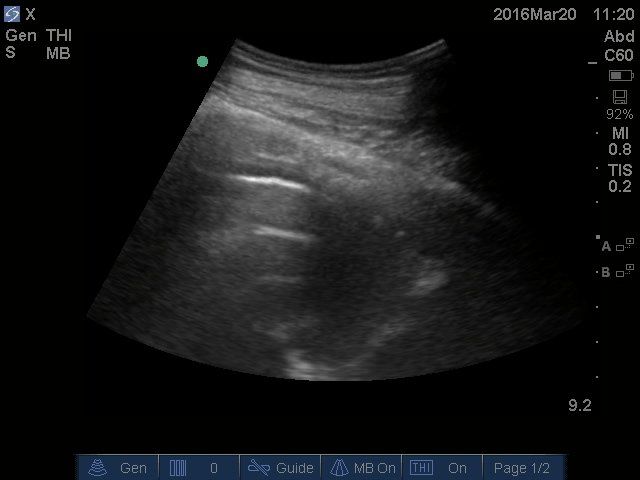
4. Hydropneumothorax
Here is a peculiar image of the thoracic cavity taken at RZ6. It appears to have two segments. To the right, there is an effusion present as demonstrated by the anechoic region with the atelectatic lung just beneath it. To the left, there is the pleural line with two A lines reverberating below.
This is a snap shot of the border between air and fluid of an hydropneumothorax as demonstrated by the CT scan. This was taken in the context of an empyema secondary to gas forming anerobes.
Below is another hydropneumothorax. Notice that on the left of the image, there is A-lines and to the right is an anechoic region (effusion). The two regions are sharply and acutely split.
Take Home Messages:
- E-Lines: ask if they are arising ABOVE the pleural line - use the Bat Wing Sign as your anchor
- Lung Pulse: it NEVER spans above the Pleural Line on M-Mode
- Investigate with formal radiological imaging modalities if you are not sure of what you are seeing
- Scan patients that have known pathologies that will yield these confounding sonographic features