
IVC - Image Acquisition
FOCUS Continuum - Image Acquisition
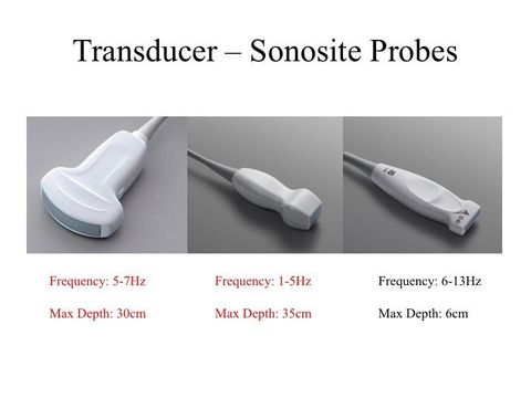
a. Probe Selection
Given the depth of the IVC, the phased array probe and curvilinear probe are ideal.
I prefer the curvilinear probe given its wide field of view - particularly when imaging the IVC from right zone 4 of the thorax.
Given the depth of the IVC, the phased array probe and curvilinear probe are ideal.
I prefer the curvilinear probe given its wide field of view - particularly when imaging the IVC from right zone 4 of the thorax.
b. Preset Optimization
Select "Cardiac" or "Abdominal" Mode.
Given that it is a deep structure, reducing the Frequency may aid with the resolution.
Select "Cardiac" or "Abdominal" Mode.
Given that it is a deep structure, reducing the Frequency may aid with the resolution.

2. Patient Positioning
There are two are sonographic windows:
a. Right upper quadrant - subcostal view
b. Thoracic right zone 4
For either sonographic windows, the ideal image easier to obtain when the patient is laying supine and flat. The IVC can be imaged when the patient is sitting on an acute angle or completely upright, but you will notice that the imaged vessel will tend to be tilted on angle and more difficult to view its union with the right atrium.
3. Probe Positioning
a. Right Upper Quadrant - Subcostal View
Grip the probe like a pen.
From the subcostal view, center the right atrium, then rotate the probe counterclockwise by ~90 degrees with the indicator pointing at ~12 o’clock. The tail of the probe is usually pointing upwards -- almost perpendicular to the abdomen.
The ultrasound beam is to cut the IVC long the longitudinal plane in its widest diameter.
Take Home Messages:
- Use the phased-array or curivilinear probe
- Consider setting a lower Frequency
- Ideally, lay the patient flat on the bed for optimal visualization