
Pleural Effusion and Thoracentesis - Anatomy Review
Anatomy Review
1. Surface Anatomy
2. Pleural Relationships
Knowledge of the anatomy will not only help you understand where you should place the probe, but, also, help you identify structures that should or should not be present.
2. Pleural Relationships
1. Surface Anatomy
You cannot PoCUS without any knowledge of surface anatomy. Otherwise, you would not know where to start scanning, and what you would be expecting to see.
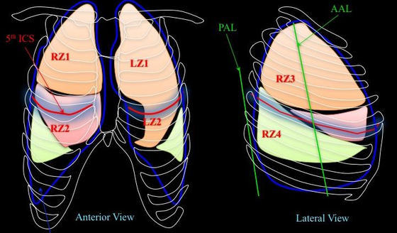
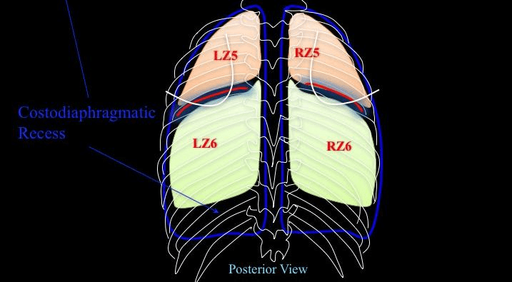
The schematic surface anatomy diaphragms shows where the parietal pleura reflects (blue line). As you can see, there is parietal pleural redundancy at the inferior aspects of the thorax - creating costodiaphragmatic recess. These are the regions that pleural fluid first gravitate towards if it is not loculated.
The most inferior recess is at the posterior aspect followed the lateral recess. Finally, the anterior aspect, also, have a recess though if you are seeing an effusion there, it either is loculated or very large.
You cannot PoCUS without any knowledge of surface anatomy. Otherwise, you would not know where to start scanning, and what you would be expecting to see.
The schematic surface anatomy diaphragms shows where the parietal pleura reflects (blue line). As you can see, there is parietal pleural redundancy at the inferior aspects of the thorax - creating costodiaphragmatic recess. These are the regions that pleural fluid first gravitate towards if it is not loculated.
The most inferior recess is at the posterior aspect followed the lateral recess. Finally, the anterior aspect, also, have a recess though if you are seeing an effusion there, it either is loculated or very large.
This table summarizes the average anatomical demarcation of the lung and parietal pleural margins in relation to surface anatomy.

2. Pleural Relationships:
Notice how the pleural space wraps around the lungs bilaterally, and their relationship to the spine.
The importance of spine is related to the Spine Sign which was first introduced in the Thoracic US - Basics module. We will be revisiting this finding in the module as it is very useful for detecting a pleural effusion.

Take Home Messages:
- VPPI is the basis of thoracic PoCUS
- Pleural recess is present not only posterior, but, also, anteriorly and laterally
- Pleural space is adjacent to mediastinal structures and the spine