
Peripheral Venous Access - Anatomical Considerations
Anatomical Considerations
Before we can use PoCUS to help us identify veins, we need to have a idea where they are, or how to find them. Unlike arteries, the location and trajectory of peripheral veins are much more variable - particularly the small branches and tributaries. However, the anatomical locations of the main veins they drain into are relatively predictable. As a result, we can use these constants to help us trace these small veins for cannulation.
Before we can use PoCUS to help us identify veins, we need to have a idea where they are, or how to find them. Unlike arteries, the location and trajectory of peripheral veins are much more variable - particularly the small branches and tributaries. However, the anatomical locations of the main veins they drain into are relatively predictable. As a result, we can use these constants to help us trace these small veins for cannulation.
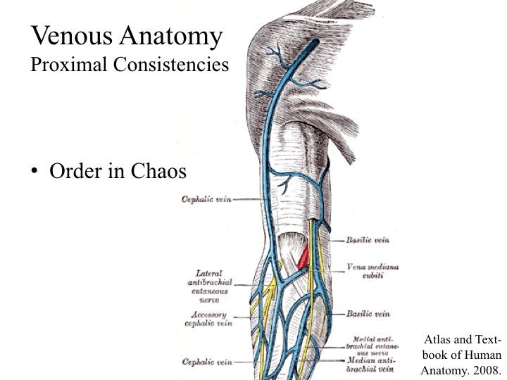
Venous Anatomy of the Proximal Upper Extremity
The main SUPERFICIAL venous branches in this region of the arm:
A pair of brachial veins that are situated adjacent to the brachial artery. They are DEEP veins of the arm. DO NOT CANNULATE THESE. (They are quite small as well.)
If a clot forms in the deep vein, a deep vein thrombosis (DVT) has been precipitated. Iatrogenically, 3 months of therapeutic anticoagulation will be warranted.
The main SUPERFICIAL venous branches in this region of the arm:
- Cephalic Vein
- Basilic Vein
- Antecubital Vein
A pair of brachial veins that are situated adjacent to the brachial artery. They are DEEP veins of the arm. DO NOT CANNULATE THESE. (They are quite small as well.)
If a clot forms in the deep vein, a deep vein thrombosis (DVT) has been precipitated. Iatrogenically, 3 months of therapeutic anticoagulation will be warranted.
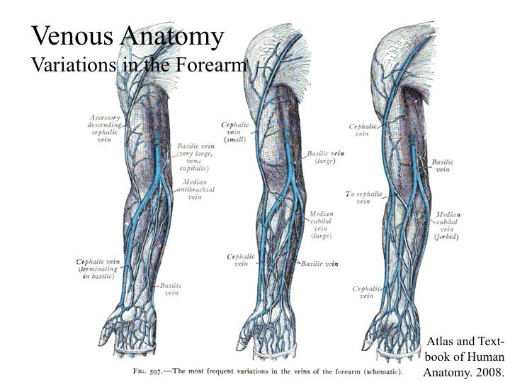
Venous Anatomy of the Distal Upper Extremity
The SUPERFICIAL venous network in this region is MUCH more variable. However, one can trace them out by first landmarking a known proximal arm vein, then tracing it distally into the forearm.
DEEP veins of the forearms: DO NOT CANNULATE
The SUPERFICIAL venous network in this region is MUCH more variable. However, one can trace them out by first landmarking a known proximal arm vein, then tracing it distally into the forearm.
DEEP veins of the forearms: DO NOT CANNULATE
- Radial veins
- Ulnar veins
- Interosseous veins
Key Message:
- Locate the small and superfical branches for cannulation by tracing along the main superificial veins to find them
- DO NOT cannulate the deep veins