
Cardiac FOCUS - Image Acquisition
a. Probe Selection
For cardiac imaging, the phased array probe is most ideal given its foot print, depth penetration, and temporal resolution.
The curvilinear probe can, also, produce cardiac images though it is quite limited due to its poor temporal resolution capability. However, it suffices to assess the IVC and for pericardial effusion - particularly at the subcostal window.

b. Preset Optimization
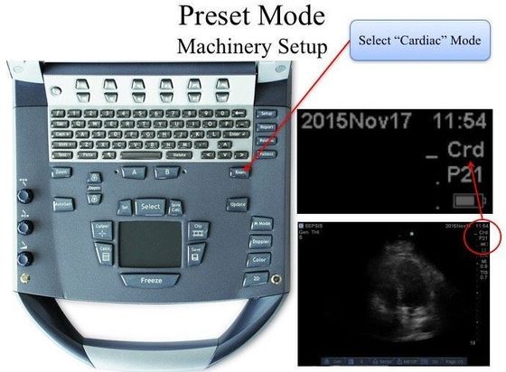
Most contemporary PoCUS machines have pre-defined settings that optimizes image quality for a particular type of scan. In this case, the interest is cardiac image acquisition, therefore the "Cardiac" Mode is selected.
Each proprietary machinery has its own methods to switch such Preset Modes. Additional tweaking can be made thereafter.
c. Indicator Position
For cardiac imaging, by convention the Emergency Medicine discipline's screen indicator is located on the LEFT whereas for Cardiology, Critical Care, Internal Medicine's screen indicator is located on the RIGHT. It really does not matter which side it is on SO LONG AS you can re-orientate your perspective lest interpretive error.
Also, REVERT the indicator position if you have changed it (eg. if you are using the EM's machine and changed the indicator to the RIGHT for a cardiac PoCUS, as a courtesy to your Emergency Physician colleagues revert it back to the LEFT after your scan).
Indicator at the Upper Left
Indicator at the Upper Right
2. Patient Position
Often, good cardiac sonographic windows can be found when the patient is laying supine and flat on the bed. However, if that is not the case, patient positioning is really the KEY MANEUVER to attain a better sonographic window for image optimization. (If the window is bad, no machine can give you good images -- it is just the limitation of physics.)
Aim:
- moves the heart CLOSER to the chest wall
- displacing the lung from a potential ultrasound window between the ribs
Maneuvers:
- left lateral decubitus: you do not have to turn the patient all the way to the left -- even by just 20-30 degrees, you will notice a difference in the image quality.
- respiration: the purpose is move the heart (or displace the lung) with respiration in hope to create a sonographic window. Ask the patient to take a SMALL breath sequentially as you evaluate for a window.
Take Home Messages:
- Use the cardiac probe
- Set the Preset to Cardiac Mode
- Have the Screen Indicator at the position that best fit your practice
- Maneuver the patient to create cardiac sonographic windows -- this is often underrated